

Auteur : Peter Forsythe – Avril 2013
BVM&S DVD MRCVS
A three-year-old, male neutered, chocolate Labrador Retriever
History
The dog was presented with a two-week history of otitis externa. There was no previous history of otitis but the dog had presented with ventral pyoderma three months previously. Further questioning revealed that the dog had also been pruritic over the feet, limbs and perineum.
Examination
General physical examination was unremarkable. Examination of the skin and ears revealed marked erythema
of both external ear canals with a bilateral, light brown coloured ceruminous aural discharge (Figure 1). The dog tolerated otoscopic examination, which revealed mild cerumenal gland hyperplasia (visible as a whitish coloured papular eruption) and an accumulation of cerumen within both ear canals, obscuring visualisation of the tympanic membranes (Figure 2). There was partial alopecia and a papular and macular eruption over the groin consistent with a pyoderma (Figure 3) and erythema over the plantar aspects of both forefeet.
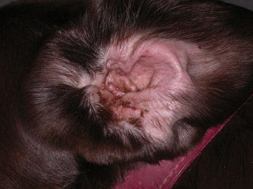
Figure 1
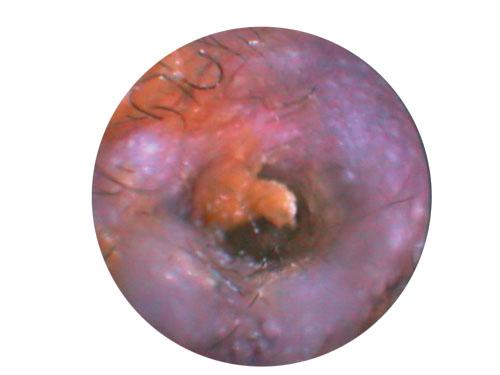
Figure 2
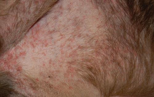
Figure 3
Problem 1
What form of otitis is described?
Discuss the relevance of the fi ndings from the examination. What would be your initial approach to this case?
Solution
This would be considered sub-acute otitis externa. Acute otitis externa is defi ned as disease being present for up to 7 days; sub-acute disease for between 7 and 30 days; and chronic disease is where disease has been active for greater than 30 days.
The description and photographs are consistent with erythematoceruminous otitis externa. In these cases, there is erythema of the pinna and /or the external ear canal, with an excessive accumulation of cerumen. The accumulation of cerumen within the ear canal would indicate a failure of epithelial cell migration – the self-cleaning mechanism that operates in a healthy ear. The accumulation of cerumen predisposes to further microbial infection and prevents penetration of topical medication. A variety of microorganisms may be associated with erythematoceruminous otitis, including Malassezia, Staphylococci and rod bacteria.
In any case of otitis, it is also important to examine the remainder of the integument as this may give clues as to
the primary cause of inflammation present. In this case, the presence of pedal pruritus and recurrent ventral pyoderma in a young Labrador Retriever were very suggestive of atopic dermatitis, the most common primary cause of inflammation in otitis externa. The pedal pruritus could also be the result of Malassezia pododermatitis. The initial approach to any case of otitis externa is to identify what, if any, infectious agents are involved, using cytology; thoroughly clean and examine the ears; and proceed with appropriate therapy for infection.
Further investigations – cytology
Samples were taken for cytology from the ventral pyoderma lesions that revealed neutrophils and intracellular cocci. Samples were also taken from both ears for cytological examination by inserting a gloved finger into the vertical ear canal. This method allows assessment of the auricular cartilage and is usually well tolerated, even when the ears are acutely inflamed. Insertion of a gloved finger is the method preferred by the author but insertion of a cotton bud into the vertical canal is a more commonly used method to collect samples for cytology.
A photomicrograph of the cytology is shown in Figure 4.
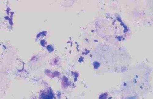
Figure 4
Problem 2
What can you identify in the photomicrograph in Figure 4?
Solution
Squames and numerous Malassezia pachydermatis yeasts can be seen from the cytology, indicating the presence of Malassezia otitis.
Treatment
A tape strip preparation from the interdigital skin failed to reveal the presence of Malassezia organisms. The dog was admitted and both ears were thoroughly cleaned under general anaesthesia. General anaesthesia is generally required to perform retrograde flushing of the ear canals, which is the only effective way of removing all discharge. Initially, a ceruminolytic product was applied to both ears and allowed to soak for a few minutes before using saline and a retrograde flushing technique. The use of video-otoscopy, as in this case, greatly facilitates visualization of the ear canals. Flushing was continued until both ear canals were thoroughly cleaned (Figure 5). This revealed the presence of intact tympanic membranes in both ears.
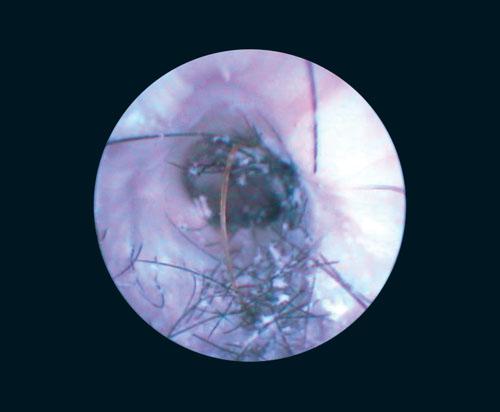
Figure 5
Problem 3
What further treatment is indicated in this case and for how long should it be continued?
Solution
There was no significant ear canal stenosis and no evidence of otitis media and, therefore, no indication for systemic antimicrobial therapy for the otitis. A ceruminolytic ear cleaner was dispensed (CleanAural®) and the owner was carefully instructed on how to perform manual ear cleaning. This instruction is essential as ear cleaning can be a difficult technique for some owners to master. Following insertion of the ear cleaner, they should be instructed on how to massage the palpable vertical ear canal for at least 30 seconds before wiping away discharge from the external portion of the vertical canal. Cotton buds should not be inserted into the canal as this can impact material deeper into the ear canal. Following ear cleaning, an antimicrobial and anti-inflammatory preparation (Canaural®) was inserted into both ears and, again, 12 hours later.
There was a pyoderma requiring treatment and clindamycin was prescribed for three weeks at a dosage of 5.5 mg/kg BID. Clindamycin was selected as it is a narrow-spectrum antibiotic generally effective against the organisms identified on cytology (most commonly Staphylococcus pseudintermedius in canine pyoderma). Ideally, a narrow-spectrum antibacterial should be used wherever possible to limit the excessive use of valuable broad-spectrum antibiotics such as cephalosporins and penicillins at a time of increasing emergence of resistant bacteria. When treating superficial pyoderma, treatment should be continued until a week after resolution of clinical signs.
Topical aural antimicrobial therapy should be continued until there is no further cytological evidence of infection. Ear cleaning should be continued until there is return to normal function of epithelial cell migration and the ear canals are able to keep themselves clean. In more severely affected cases, this function may never return and lifelong ear cleaning is required.
After treatment with CleanAural and Canaural
At a recheck after 14 days of therapy, the otitis and aural pruritus had resolved. Repeat cytology failed to reveal the presence of infection. The ventral pyoderma lesions had resolved.
Systemic antibiotic treatment was continued for a further week. The Canaural was withdrawn but ear cleaning was continued on a twice weekly basis.
A further recheck was performed after another two weeks. At that time, the owner reported that the dog was still pruritic over the feet and there had been an increase in aural pruritus.
Examination revealed erythema of the external ear canal and interdigital skin but no evidence of infection on cytology.
The ventral pyoderma had resolved.
Problem 4
Why has the aural pruritus recurred? What would you do now?
Solution
The recurrence of aural pruritus is likely to be due to withdrawal of the topical glucocorticoid therapy in the Canaural. The ongoing aural and pedal pruritus and erythema were consistent with atopic dermatitis. This is one of the most common primary causes of inflammation in otitis externa. Control of atopic dermatitis is an essential component in preventing repeated episodes of otitis externa and the problems associated with chronic otitic disease such as resistant infections and ear canal stenosis.
The initial focus is on trying to identify whether food and/ or environmental allergens are involved. The dog was started on a proprietary novel protein diet and all other food sources were excluded, including chews, biscuits,
treats, flavoured toys and toothpaste. Twice-weekly CleanAural ear cleaning was continued during the restricted diet trial. A week after starting the diet, an intradermal test was performed that gave positive reactions to house dust and storage mites. Following intradermal testing, pruritus was controlled with intermittent use of systemic glucocorticoid therapy, using prednisolone at a dosage of 0.5mg/kg SID given daily for three days and repeated as required. This was sufficient to control pruritus without masking any response to the diet trial. After eight weeks of the diet trial, there had been no further episodes of pyoderma or Malassezia otitis but aural pruritus and
erythema were still evident and there was still a requirement for intermittent glucocorticoid therapy to control pruritus.
This confirmed a diagnosis of atopic dermatitis and excluded the involvement of food allergens.
Problem 5
What are the options for long-term management in this case?
Solution
Possible options for controlling atopic dermatitis include allergen avoidance and allergen-specific immunotherapy; control of secondary infection and ongoing ear cleaning; pharmacotherapy to control pruritus, including antihistamines, essential fatty acids, Chinese herbal therapy, glucocorticoids and ciclosporin.
The dog was started on allergen-specific immunotherapy. The owner was advised to continue twice-weekly ear cleaning. Intermittent glucocorticoid therapy was continued for the first four months of allergen-specific immunotherapy to control aural and pedal pruritus.
Long-term follow-up a year later revealed that the dog had a further episode of otitis three weeks after ear cleaning was withdrawn by the owner because of a lack of symptoms. The episode of otitis responded to further topical antimicrobial therapy. There had also been a further episode of pyoderma along with Malassezia pododermatitis that responded to systemic antibacterial therapy and 2% chlorhexidine/ 2% miconazole shampoo (Malaseb® Shampoo). Ongoing weekly Malaseb baths, twice-weekly ear cleaning and allergenspecific immunotherapy had been sufficient to prevent further symptoms. There was no longer a requirement for glucocorticoid therapy. The episode of otitis following withdrawal of ear cleaning convinced the owner of the necessity for ongoing treatment.
