
Auteur : Richard Halliwell – Février 2013
MA, VetMB, PhD, DACVD, FMedSci, MRCVS
Key points
- There are striking similarities between atopic dermatitis in humans and in dogs.
- Although IgE is still thought to play a major role in the release of mast cell–derived mediators and also in allergen capture, the immunopathogenesis of canine atopic dermatitis (CAD) is now known to be far more complex. In the past, a simplistic view was generally held on the pathogenesis of CAD, with a major role for IgE and mast cells and their mediators.
- Evidence has shown that the major route of allergen access in CAD is percutaneous, although administration of allergen via the respiratory and oral routes can also exacerbate pruritus in sensitized dogs, albeit less efficiently.
- Overproduction of IgE is typically associated with a Th2 response and thus it is relevant to assess the extent to which the cellular response in CAD is Th1 or Th2 driven. Early lesions are thought to be associated with a predominantly Th2 response which changes, due to the ensuing chronicity and secondary infection, to a Th1 response.
- Cytokines derived from keratinocytes, T lymphocytes, and macrophages, many of which are pro-inflammatory, are present in the inflammatory milieu in lesional skin of dogs with CAD. However, there is a paucity of definitive information implicating specific mediators in the dog, and further carefully planned studies are urgently needed.
- Dermatologists treating human patients with atopic dermatitis (AD) have long believed that barrier function abnormalities play a crucial role in the pathogenesis. These defects appear to play a role in dogs as well, as assessed by measurement of filaggrin, transepidermal water loss, analysis of surface lipids such as ceramides, and ultrastructural studies showing differences between atopic and normal dogs. The extent to which this contributes to the pathogenesis, or results from inflammation, is unknown.
- Atopic dermatitis has recently been redefined by the International Task Force on Canine Atopic Dermatitis as “a genetically predisposed inflammatory and pruritic skin disease with characteristic clinical features associated with IgE antibodies most commonly to environmental allergens.”
- In around 10% of classical cases of CAD, no relevant allergen-specific IgE is demonstrable. Such cases are termed atopic-like dermatitis. The optimal therapy for this frustrating condition is not known.
- Many experienced clinical veterinary dermatologists still make the diagnosis of CAD by 1) observation of compatible clinical signs and 2) failure to document any explanation for these signs even though three sets of diagnostic criteria have been proposed
The atopic diseases in humans
The term atopy, derived from the Greek and literally translated as “strange disease,” was introduced by Coca and Cooke in the 1920s to embrace asthma and hay-fever which are familial hypersensitivity disorders of humans.1 They noted that the conditions were associated with an unusual antibody which they termed reagin, which was 1) heat labile, and 2) could be transferred to the skin of normal individuals by the so-called Prausnitz-Küstner (or PK) test.2 Many years later, the painstaking work of the Ishizakas and their colleagues determined that this antibody belonged to a hitherto-undescribed antibody class, which was named immunoglobulin E (IgE).3 Atopic dermatitis (AD), another familial disease, was added to this group of diseases later by Hill and Sulzberger,4 and through the ages, affected patients have often exhibited “the atopic march,” where their disease commences with AD and they later develop asthma or hay-fever. Although AD and asthma are usually associated with excessive production of IgE and exacerbated by environmental allergens, a subset exists, namely “intrinsic,” in which allergen-specific IgE does not appear to be involved.
Eczema and atopic dermatitis in dogs
One of the early classical descriptions of “eczema” in dogs was in a paper by Schnelle working at the Angell Memorial Hospital in Boston. He reported that 15% of all cases seen at his clinic were accorded a diagnosis of “eczema” and that 56.9% of all dogs with skin disease and 26.6% of all cats similarly affected were deemed to be suffering from this condition.5 Comparable data were reported from the clinics at Cornell University in Ithaca, New York.(6)
Although it was generally believed that “eczema” was a manifestation of allergy, the exact nature of the inciting cause was controversial, with most emphasis being placed upon foods. In addition, flea allergy was recognized as an important cause of “summer eczema.”(7) The first documentation that environmental allergens might also be involved was provided in 1941 by Whittich, a human allergist, who gave an elegant description of a dog with perennial pruritus due to a food allergy that suffered from seasonal hay-fever due to a concomitant pollen allergy.8 The dog was treated with an appropriate hypoallergenic diet and successfully hyposensitized with injections of allergenic extracts of the pollens to which sensitivity was shown. The association with IgE was further confirmed by demonstrating positive PK tests using both canine and human recipients.
Little progress was made until the 1960s when investigations were undertaken on canine ragweed pollenosis in the United States by Roy Patterson, another human physician. He proposed that dogs suffering from this condition could represent a good model for hay-fever and allergic rhinitis of humans, and he developed a colony of atopic dogs suffering from the condition.9 The dogs were reported as showing signs of hay-fever, and although they did not suffer from spontaneous asthma, the latter was inducible by insufflation with high concentrations of allergen. Furthermore, asthma was inducible in normal dogs following injection of serum from allergic dogs and subsequent antigenic challenge.10 Despite the fact that there were obvious dermatologic signs in addition to hay-fever-like signs, it was not thought to be truly analogous to AD of humans. Instead it was termed atopy, atopic disease, or allergic inhalant dermatitis, the latter term in the mistaken belief that inhalation was the major route of access of allergen.
This period saw the first detailed clinical descriptions of the condition as seen in clinical veterinary practice.(11) “Atopy” was described as a familial, pruritic dermatitis with a predominantly facial and ventral distribution, often presenting initially without primary lesions, but with chronic changes developing in association with self-trauma. Some patients presented also with hay-fever like signs and rubbed at their nasal and ocular region with or without accompanying dermatitis. Patients presented with seasonal or perennial signs, with many cases commencing seasonally and progressing to perennial involvement.
The pathogenesis of canine atopic dermatitis
Characterization of Canine IgE
Classically, allergic asthma and hay-fever of humans were believed to be associated with IgE antibody, although its role in AD has always been more controversial. As the initial view of canine “atopy” or “atopic disease” was a condition not truly analogous to AD of humans, the major emphasis of early work was on the characterization of canine IgE as a probable key player in the immunopathogenesis. The identification and description of canine IgE was reported in 1973 only 6 years after that of its human counterpart,12 with which it was antigenically similar, and it was shown to be localized on mast cells in canine skin.(13) The development of tests for the measurement of canine allergen-specific IgE followed shortly therafter.(14)
What is the Role of IgE in the Pathogenesis of CAD?
In the 1970s and 1980s a simplistic view was generally held on the pathogenesis of CAD with a major role for IgE and mast cells and their mediators. Although IgE is still thought to play a major role in the release of mast cell–derived mediators and also in allergen capture, the immunopathogenesis is now known to be far more complex.
Allergen Access, Capture, and Processing
The term allergic inhalant dermatitis— which was in common use in the 1980s— implied on the basis of mere supposition that the route of access of allergen was via inhalation. Evidence in favor of the percutaneous route was derived from the work of Olivry, who showed that lesions skin of dogs with CAD showed focal proliferation of Langerhans cells and that these cells were armed with IgE antibody.(15) There was also proliferation of dermal dendritic cells also armed with IgE, which implied that these cells were involved in allergen processing.16 It was only in 2006, however, that Marsella, employing the high IgE-producing beagle model of AD, provided direct evidence that the major route of allergen access was percutaneous, although administration of allergen via the respiratory and oral routes could also exacerbate pruritus in sensitized dogs albeit less efficiently.(17)
Inflammatory Cells in CAD – Are They Indicative of a Th1 or Th2 Response?
Immunohistochemical studies have shown that the infiltrating cells in skin biopsies of spontaneous cases of CAD comprise mast cells, dendritic antigen-presenting cells, T lymphocytes expressing ?d rather than aß receptors with low numbers of neutrophils and eosinophils and rare B-lymphocytes.16 Both CD4+ and CD8+ T cells are found in increased numbers, with a major increase in CD8+ cells in the epidermis along with microaggregates of eosinophils.16 Classically, T cells can be assigned on the basis of function to Th1, promoting cell-mediated immunity, and Th2 favoring antibody production, including IgE. Thus overproduction of IgE is typically associated with a Th2 response. It is pertinent, therefore, to assess the extent to which the cellular response in CAD is Th1 or Th2 driven.
Studies employing a non-quantitative reverse transcriptase polymerase chain reaction (PCR) on clinical cases suggested that a clear Th2 polarization was evident in some 25%.18 A later study also employing clinical material using semi-quantitative methods yielded evidence of overexpression of both Th1 (interferon gamma, IFN-?) and Th2 (interleukin-4, IL-4) cytokines.(19,20) It was suggested that early lesions might be associated with a predominantly Th2 response which changed due to the ensuing chronicity and secondary infection to a Th1 response, conclusions supported by the most recent publication on this topic.(21)
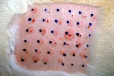
The Th2 versus Th1 issue was investigated further using atopy patch tests in the high-IgE beagle model in which it should be possible to separate out the acute and chronic phases. Among the Th2 cytokines, IL-6 and IL-13 were significantly increased and peaked at 24 hours.22 Although IL-4 increased over 6 to 24 hours, the increase was not significant. Among the Th1 cytokines, IFN-? had a biphasic response with peaks at 6 hours and 96 hours with IL-18 gradually increasing through 96 hours. Thus although there is a pattern which is in general accord with that in humans, results are not conclusive and further studies are required.
The issue has also been investigated using whole blood from clinical cases. The first of these assessed mRNA of IFN-?, IL-4, IL-5, and IL-10 in freshly isolated peripheral blood mononuclear cells (PBMCs) from clinical cases.23 The results were inconclusive, with a reduction in IFN-? and an increase in IL-5, with no change in IL-4 and IL-10. The second study evaluated mRNA expression of IL-4, IL-13, IL-10, and transforming growth factor beta (TGFß) in the high-IgE beagle model. IL-4 and IL-13 were unchanged, but the levels of expression of the immunosuppressive cytokines IL-10 and TGFß were reduced, which could imply aberrant regulatory T-cell function.(24)
Interest has also focused on the possible role of CC chemokines in CAD. Thymus and activation regulated chemokine (TARC) is produced mainly from keratinocytes in response to inflammatory cytokines such as IL-1ß, IFN-?, and tumor necrosis factor alpha (TNF-a). It plays an important role in Th2 cell migration since its receptor (CCR4) is expressed selectively on Th2 cells. TARC was found to be expressed exclusively on lesional skin of atopic dogs, and was indeed associated with increased expression levels of IL-1ß, IFN-?, and TNF-a.25 In this study of chronic clinical CAD, no increase in IL-4 was detectable. A later study by the same workers employing a monoclonal antibody to TARC confirmed keratinocytes in lesional skin of CAD as the major source, and that its receptor (CCR4) was expressed on the infiltrating cells.(26) The importance of TARC was also derived from studies using patch tests on the high IgE-producing beagle model.(22) In another study of seven chemokines, levels of the CCL28 expression in lesional skin was significantly increased, whereas those of CCL27 were significantly reduced.(27)
What Are the Mediators of Inflammation and Pruritus in CAD?
Implication of any specific mediator(s) in the development of inflammation and pruritus in CAD is exceedingly difficult. Ideally, studies should show 1) evidence of increased levels in the skin, 2) development of pruritus and inflammation following intradermal injection in normal animals, and 3) amelioration of signs following specific inhibition.
The role of histamine has long been controversial. Histamine levels have been reported elevated in the skin of dogs with AD(28) and histamine is released from basophils of atopic dogs in response to allergen.29 The generally poor response to antihistamine therapy, however, and negative results in a study employing the Maltese beagle cross model of CAD precludes a major role for this mediator.30 Interest has also focused on other mast cell–derived mediators including proteases and leukotrienes (LT). In one study, elevated levels of LTB4 were found in a number of inflammatory skin diseases including CAD31, but the significance of sulfido-LT was later questioned by equivocal results using the high IgE-producing beagle model.(32)
There are, of course, a whole plethora of cytokines derived from keratinocytes, T cells, and macrophages present in the inflammatory milieu in lesional skin of dogs with CAD, many of which are pro-inflammatory. Great interest in the study of human AD is the role of IL-31, with levels in sera of patients with AD correlate closely with the level of pruritus and with total IgE.33 Interestingly, however, there were no significant differences between patients with extrinsic (allergic) and intrinsic AD. There is interest also in the role of the calcium binding protein A8 (S100A8), and a recent study showed higher levels in CAD patients and a correlation with disease severity.34 Elevated levels of this protein are not exclusive to CAD, but may be seen on other inflammatory skin diseases. Finally, the possible role of neuro-immune reactions must be mentioned and neuropeptides and neurotropins, although veterinary studies supporting their involvement are lacking.
In summary, there is a paucity of definitive information implicating specific mediators in the dog, and further carefully planned studies are urgently needed.
Is Barrier Function Defective in CAD?
Filaggrin
Dermatologists treating human patients with AD have long believed that barrier function abnormalities play a crucial role in the pathogenesis, and placed great emphasis on restoration of function as a major therapeutic target. Of great importance was the recent discovery of a genetically controlled abnormality in the structural protein filaggrin in many human patients with AD.(35) Immunofluorescent studies in dogs first showed that filaggrin staining differed between atopic and normal dogs with finer granules and less intensity of staining in the former.36 Of great interest was the observation that atopic dogs showed less staining than did normal dogs, and that the staining of normal skin was reduced after dust mite exposure.37 Very recently a loss-of-function mutation was identified in 4 of 18 atopic dogs analyzed, once again highlighting the similarities between human and canine AD.(38)
Transepidermal Water Loss
The integrity of barrier function is generally assessed by measurement of transepidermal water loss (TEWL). Although some doubt that this provides an accurate assessment,(39) the relationship between the two in dogs has recently been confirmed using tape stripping and gauging the barrier function by permeation of a fluorescent dye.(40) TEWL measurement gives differing results, however, depending on the precise technique used (41,42,43) (open versus closed chamber, site variations, movement and presence or absence of hair) and careful validation is necessary. Despite these reservations, some important data has emerged from the beagle model confirming that TEWL is increased in sites prone to the development of CAD prior to allergen exposure, and this is further increased in diseased skin when compared with age-matched normal beagles.(44)
Analysis of Surface Lipids
Ceramides play a major role in maintaining barrier function, and where they are decreased, a vicious cycle can ensue wherein bacterial colonization that is a feature of AD can lead to further lowering of epidermal ceramide levels through action of bacterial ceraminidases.(45) A recent study has shown that the surface lipids of non-lesional skin of dogs with AD do indeed differ from those of normal dogs.(46) The levels of ceramides 1 and 9 were significantly decreased, whereas that of cholesterol was significantly increased, and ceramide/cholesterol ratio was significantly lower. The changes in ceramide 1 may be of especial significance, as this lipid is believed to be of particular importance in the assembly of the intercellular lipid lamellae.(47) A more recent study has confirmed that ceramides are reduced in both lesional and non-lesional skin of atopic dogs, and that this reduction is inversely correlated with the transepidermal water loss.(48) More recently, 11 clusters of peaks representing free ceramide classes have been demonstrated, similar to those seen in humans, and the same three classes known to be reduced in human AD were found similarly reduced in CAD.(49) Sphingosines, which can be cleaved from ceramides, are also important and have marked protective properties. A recent study demonstrated significantly lower levels of sphingosine-1-phosphate in skin from atopic canines as compared with normals, implying an imbalance in the S1P-S1P-lysase axis.(50)
Ultrastructural Studies
Three ultrastructural studies have reported similar findings in dogs with AD.(51,52,37) Instead of being organized into lamellae, the lipid deposits are reduced in both lesional and non-lesional skin and the deposits are heterogeneous, with widened intercellular spaces. In one study, delayed release of lamellar bodies was noted, and there was a sudden release of lamellar lipids upon allergen challenge.(37)
In summary, multiple studies from varying angles all point to abnormalities in barrier function and these are obvious therapeutic targets.
The clinical features of cad
Definitions
Atopic dermatitis has recently been redefined by the International Task Force on Canine Atopic Dermatitis53 as:
“A genetically predisposed inflammatory and pruritic skin disease with characteristic clinical features associated with IgE antibodies most commonly to environmental allergens.”
In some cases (around 10%) of classical CAD, no relevant allergen-specific IgE is demonstrable. Such cases are termed atopic-like dermatitis. This is exactly parallel to the situation in humans where some patients suffer from “intrinsic AD,” where there is no evidence of allergen-specific IgE. The definition of atopic-like dermatitis is thus:
“A genetically predisposed inflammatory and pruritic skin disease with clinical features identical to those of atopic dermatitis in which IgE antibodies to environmental allergens are not demonstrable.”
Breed Predilections
Breed predilections have long been recognized, and the mode of inheritance is currently under investigation. Those breeds shown to be significantly predisposed in two recent studies were Labrador retriever,(54) golden retriever,(54) West Highland White terrier,(54,55) Chinese shar-pei,(54) bull terrier,(54,55) Bichon Frisé,(54) Tibetan terrier,(54) English springer,(54) Boxer,(55) French bulldog,(55) Dalmatian,(55) Hungarian Vizsla,(55) and Basset hound.(5)
Age of Onset
Canine atopic dermatitis is a disease that commences in the young dog, with 78% showing clinical signs at less than 3 years and 16% at less than 1 year.(55) It is exceedingly rare for signs to commence in dogs older than 7 years of age.(11)
The Primary Disease
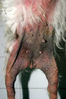
The cardinal sign is itching, and in the early stage there may be little evidence of inflammation. Later on, erythematous macules and papules develop. The distribution is predominantly ventral and facial, with pedal involvement almost invariably seen. Typically, the flexural surfaces of the limbs are involved. The distribution of clinical signs is at least in part reflective of the route of access of allergen. Otitis is very common, and probably affects all cases at some point in their course. Occasionally it may be unilateral. The otitis associated with CAD in the early stages is highly characteristic and involves the inner ear flap and the vertical canal. When the horizontal canal is observed, it appears normal in the early case although it becomes secondarily infected in chronic cases, with accompanying secondary pathologic changes. In some cases otitis is the only sign. There is some evidence of breed variability in the presenting signs.(56)
There exists much controversy regarding the possible involvement of other body systems. Although asthma can be induced in susceptible animals using insufflation of high concentrations of allergen, it is rare to non-existent in clinical CAD. This may be a reflection of the variations in regional mast cell density between humans, the cat (where allergic asthma does occur (57)), and dogs. Certainly some animals appear to have concomitant hay-fever-like signs, and a recent publication has highlighted the importance of allergic conjunctivitis, which the authors claim is often overlooked.(58) Although the existence of an allergic tracheobronchitis as a manifestation of atopic disease has been proposed, convincing documentation is lacking.
Secondary Complications
In time, evidence of self-trauma, hair loss, and seborrheic changes develop. There may be areas of hyperpigmentation, lichenification, and crusting. The otitis often worsens, and progressive pathologic changes lead to hypertrophy of the lining and consequent narrowing of the ear canal. Sometimes a verrucose proliferation develops which can largely obstruct the canal. Atopic dogs are predisposed to bacterial overgrowth and then to overt pyoderma. This is usually a folliculitis, but occasionally some cases develop a deep pyoderma. The atopic dog is, in fact, “a pyoderma waiting to happen” because:
- Corneocytes from atopic dogs show enhanced adherence to staphylococci.(59)
Staphylococci show greater adherence to corneocytes from inflamed skin.(60)
Adherence has been shown to correlate with pruritus scores61 although in this study there was no apparent correlation with the propensity to develop pyoderma.
Atopic dogs have been shown to have a significantly higher level of staphylococci at carrier sites as compared with normals.(62)
The propensity to develop anti-staphylococcal IgE has also been shown,63 thus compounding the effects.
Similarly, the ubiquitous yeast Malassezia is found in higher numbers on the skin of dogs with dermatologic diseases,(64) although precise data linking Malassezia overgrowth specifically to CAD is lacking. Nonetheless, the fact that sera from atopic dogs have higher levels of Malassezia-specific IgE emphasizes the importance of this organism as a contributor to the total allergenic load65 and to the development of Malassezia dermatitis, manifesting typically as areas of greasy erythema proceeding to lichenification in chronic cases.
Diagnostic criteria
The first paper suggesting that specific criteria would be helpful in the diagnosis of CAD was that of Willemse(66) and colleagues, which was followed by some modifications proposed by Prelaud and co-workers.67 More recently, a revised set of criteria based upon a more limited number of observations was developed by Favrot and colleagues.(68) Two sets were proposed, namely:
Set 1 |
Set 2 |
| Age at onset < 3 years | Age at onset < 3 years |
| Mostly indoor | Mostly indoor |
| Corticosteroid responsive pruritus | Pruritus sine materia at onset |
| Chronic or recurrent yeast infections | Affected front feet |
| Affected front feet | Affected ear pinnae |
| Affected ear pinnae | Non-affected ear margins |
| Non-affected ear margins | Non-affected dorso-lumbar area |
| Non-affected dorso-lumbar area |
What Do We Know About “Atopic-like” Dermatitis?
Very little work has been done on this intriguing disease entity. We know nothing about barrier function, and nor do we know about optimal therapy for this frustrating condition.
Conclusions
This paper has presented a historical overview of CAD and reviewed the current state of knowledge of the pathogenesis. The past 20 years has witnessed an explosion of research in this area, and the recognition of the striking similarities between CAD and the human counterpart will hopefully enable a more ready source of funding to support future endeavors.
References
-
- Coca AF, Cooke RA. On the classification of the phenomena of hypersensitiveness. J Immunol. 1923;8: 163-166.
- Prausnitz C, Kustner H. Studien über die Uberimfindlichkeit. Zentralb Bakt. 1921;86:160-169.
- Ishizaka K, Ishizaka T, Hornbrook MM. Physicochemical properties of human reaginic antibody. IV. Presence of a unique immunoglobulin as a carrier of reaginic activity. J Immunol. 1966; 97:75-85.
- Hill LW, Sulzburger MB. Evolution of atopic dermatitis. Arch Dermatol. 1935;32:451-455.
- Schnelle GB. Eczema in dogs – an allergy. North Am Vet. 1933;14:37-40.
- Milks HJ. The role of allergy in skin diseases of dogs. Cornell Vet. 1938;28:142-147.
- Kissileff A. The dog flea as a causative agent in summer eczema. J Am Vet Med Assoc. 1938;83:21-24.
- Wittich FW. Spontaneous allergy (atopy) in the lower animal. J Allergy. 1941;12:247-257
- Patterson R, Chang WW, Pruzansky JJ. The Northwestern Colony of Atopic Dogs. J Allergy Clin Immunol. 1963; 34:455-459.
- Patterson R, Sparks DB. The passive transfer to normal dogs of skin test reactivity, asthma and anaphylaxis from a dog with ragweed pollen hypersensitivity. J Immunol. 1962;88:262.
- Halliwell REW, Schwartzman RM. Atopic disease in the dog. Vet Rec. 1971;89:209-213.
- Halliwell REW, Schwartzman RM, Rockey LH. Antigenic relationship between human and canine IgE. Clin Exp Immunol. 1972;10:399-407.
- Halliwell REW. The localization of IgE in canine skin: an immunofluorescent study. J Immunol. 1973;110: 422-430.
- Halliwell REW, Kunkle GA. The radioallergosorbent test in the diagnosis of canine atopic disease. J Allergy Clin Immunol. 1978;62:236-244.
- Olivry T, Moore PF, Affolter VK, Naydan DK. Langerhans cell hyperplasia and IgE expression in canine atopic dermatitis. Arch Dermatol Res. 1996;288:579-585.
- Olivry T, Naydan DK, Moore PF. Characterization of the cutaneous inflammatory infiltrate in canine atopic dermatitis. Am J Dermatopath. 1997;19:477-486.2012
- Marsella R, Nicklin C, Lopez J. Studies on the route of access of allergen exposure in high IgE-producing beagkle dogs sensitized to house dust mites. Vet Dermatol. 2006;17:306-312.
- Olivry T, Dean GA, Tomkins MB, et al. Toward a canine model of atopic dermatitis: amplification of cytokine- gene transcripts in the skin of atopic dogs. Exp Dermatol. 1999;8:204-211.
- Nuttall TJ, Knight PA, McAleese SM, et al. T-helper 1, T-helper 2 and immunosuppressive cytokines in canine atopic dermatitis. Vet Immunol Immunopathol. 2002;87:379-384.
- Nuttall TJ, Knight PA, McAleese SM, et al. Expression of Th1, Th2 and immunosuppressive cytokine gene transcripts in canine atopic dermatitis. Clin Exp Allergy. 2002;32:789-795.
- Sclotter YM, Rutten VP, Riemers FM, et al. Lesional skin in atopic dogs shows a mixed Type-1 and Type- 2immune responsiveness. Vet Immunol Immunopathol. 2011;143:20-26.
- Marsella R, Olivry T, Maeda S. Cellular and cytokine kinetics after epicutaneous allergen challenge (atopy patch testing) with house dust mites in high IgE beagles. Vet Dermatol. 2006;17:111-120.
- Hayashiya S, Tani K, Morimoto M, et al. Expression of T helper 1 and T helper 2 cytokine mRNAs in freshly isolated peripheral blood mononuclear cells from dogs with atopic dermatitis. J Vet Med. 2002;49:27-31.
- Maeda S, Tsuchida H, Marsella R. Allergen challenge decreases mRNA expression of regulatory cytokines in whole blood of high-IgE beagles. Vet Dermatol. 2007;18:422-426.
- Maedea S, Fujiwara S, Omori K, et al. Lesional expression of thymus and activation-regulated chemokine in canine atopic dermatitis. Vet Immunol Immunopathol. 2002;88:79-87.
- Maeda S, Tsukui T, Saze K, et al. Production of a monoclonal antibody to canine thymus and activation- regulated chemokine (TARC) and detection of TARC in lesional skin from dogs with atopic dermatitis. Vet Immunol Immunopathol. 2005;103:83-92.
- Maeda S, Tsuchida H, Shibata S, et al. Expression analysis of CCL27 and CCL28 mRNA in lesional and non- lesional skin of dogs with atopic dermatitis. J Vet Med Sci. 2008;70:51-55.
- Helton-Rhodes K, Kerdel F, Soter NA, Chinnici R. Investigation into the pathogenesis of canine atopy. Sem Vet Med Surg (Small Animal). 1987;2:199-201.
- Jackson HA, Miller HR, Halliwell RE. Canine leukocyte histamine release in response to antigen and to anti-IgE. Vet Immunol Immunopathol. 1996;53:195-206.
- Bäumer W, Stahl J, Sander K, et al. Lack of preventing effect of systemically and topically administered H(1) or H(4) receptor antagonists in a dog model of acute atopic dermatitis. Exp Dermatol. 2011;20:577-581.
- Keitzman M. Eicosandoid levels in canine inflammatory skin disease. In Von Tscharner C, Halliwell REW (eds): Advances in Veterinary Dermatology, vol 1. London: Balliere Tindall, 1990, pp 211-220.
- Marsella R, Nicklin CF. Sulpido-leukotriene production from peripheral leukocytes and skin in clinically normal dogs and in house dust mite positive atopic dogs. Vet Dermatol. 2001;12:3-12.
- Kim S, Kim H-J, Yang HS, et al. IL-31 serum protein and tissue mRNA levels in patients with atopic dermatitis. Ann Dermatol. 2011;23:468-473.
- Chung TH, Oh JS, Lee YS, et al. Elevated serum levels of S100 calcium binding protein A8 (S100A8) reflect disease severity in canine atopic dermatitis. J Vet Med Sci. 2010;72:693-700.2012 Symposium Proceedings Allergic Skin Disease 14
- Palmer CN, Irvine AD, Terron-Kwaitkowski A, et al. Common loss of function variants of the epidermal barrier protein filaggrin are a major predisposing factor for atopic dermatitis. Nat Genet. 2006;38:441-446.
- Marsella R, Samuelson D, Harrington L. Immunohistochemical evaluation of filaggrin polyclonal antibody in atopic and normal beagles. Vet Dermatol. 2009;20:547-554.
- Marsella R, Samuelson D. Unravelling the skin barrier: a new paradigm for atopic dermatitis and house dust mites. Vet Dermatol. 2009;20:533-540.
- 38. Chervet L, Galichet A, McLean WHI, et al. Missing C-terminal filaggrin expression, NFkappaB activation and hyperproliferation identify the dog as a putative model to study epiermal dysfunction in atopic dermatitis. Exp Dermatol. 2010;19:343-346.
- Chilcott RP, Dalton CH, Emmanuel AJ, et al. Transepidermal water loss does not correlate with skin barrier function in vitro. J Invest Dermatol. 2002;118:871-875.
- Shimada K, Yoshihara T, Yamamoto M, et al. Transepidermal water loss (TEWL) reflects skin barrier function of dog. J Vet Med Sci. 2008;70:841-843.
- Watson A, Fray T, Clarke S, et al. Reliable use of the ServoMed Evaporimeter EP-2TM to assess transepidermal water loss in the canine. J Nutr. 2002;132:1661S-1664S.
- Hester SL, Rees CA, Kennis RA, et al. Evaluation of corneometry (skin hydration) and transepidermal water- loss measurements in two canine breeds. J Nutr. 2004;134:2110S-2113S.
- Lau-Gillard PJ, Hill PB, Chesney CJ, et al. Evaluation of a hand-held evaporimeter (VapoMeter®) for the measurement of transepidermal water loss in healthy dogs. Vet Dermatol. 2010;21:136-145.
- Hightower K, Marsella R, Creary E. Evaluation of trans-epidermal water loss in canine atopic dermatitis: a pilot study in beagle dogs sensitized to house dust mites. Vet Dermatol. 2008;19:108.
- Ohnishi Y, Okino N, Ito M, Imayama S. Ceramidase activity in bacterial skin flora as a possible cause of ceramide deficiency in atopic dermatitis. Clin Diag Lab Immunol. 1999;6:101-104.
- Reiter LV, Tores SMF, Wertz PW. Characterization and quantification of ceramides in the non-lesional skin of canine patients with atopic dermatitis compared with controls. Vet Dermatol. 2009;20:260-266.
- Wertz PW. Epidermal lipids. Seminars Dermatol. 1992;11:106-113.
- Shimada K, Yoon J-S, Yoshihara T, et al. Increased transepidermal water loss and decreased ceramide content in lesional and non-lesional skin of atopic dogs. Vet Dermatol. 2009;20:541-546.
- Yoon J-S, Nishifuji K, Sasaki A, et al. Alteration of stratum corneum ceramide profiles in spontaneous canine model of atopic dermatitis. Exp Dermatol. 2011;20:732-736.
- Bäumer W, Roßbach K, Mischke R, et al. Decreased concentration and enhanced metabolism of sphingosine- 1-phosphate in lesional skin of dogs with atopic dermatitis: disturbed sphingosine-1-phosphatase homeostasis in atopic dermatitis. J Invest Dermatol. 2011;131: 266-268.
- Inman AO, Olivry T, Dunston SM, et al. Electronic microscopic observations of stratum corneum intercellular lipids in normal and atopic dogs. Vet Pathol. 2001;38:720-723.
- Piekutowski A, Pin D, Reme CA, et al. Effects of a topically applied preparation of epidermal lipids on the Stratum Corneum barrier of atopic dogs. J Comp Pathol. 2008;138:197-203.
- Halliwell R. Revised nomenclature for veterinary allergy. Vet Immunol Immunopathol. 2006;114:207-208.2012 Symposium Proceedings Allergic Skin Disease 15
- Zur G , Ihrke PJ, White SD, Kass PH. Canine atopic dermatitis: A retrospective study of 226 cases examined at the University of California, Davis, 1992-1998. Part 1. Clinical features and allergy testing results. Vet Dermatol. 2002;13:89-102.
- Picco F, Zini E, Nett C, et al. A prospective study on canine atopic dermatitis and food-induced allergic dermatitis in Switzerland. Vet Dermatol. 2008;19:150-155.
- Wilhelm S, Kovalik M, Favrot C. Breed-associated phenotypes in canine atopic dermatitis. Vet Dermatol. 2010;22:143-149.
- Prost C. Treatment of feline asthma with allergen avoidance and specific immunotherapy: Experience with 20 cats. Rev Franc Allerg Immunol Clin. 2008;48:49-413.
- Lourenco-Martins AM, Delgardo E, Neto I, et al. Allergic conjunctivitis and conjunctival provocation tests in atopic dogs. Vet Ophthalmol. 2011;14:248-256.
- McEwan NA. Adherence by Staphylococcus intermedius to canine keratinocytes in atopic dermatitis. Res Vet Sci. 2000;68:279-283.
- McEwan NA, Mellor D, Kalna G. Adherence by Staphylococcus intermedius to canine corneocytes: a preliminary study comparing noninflamed and inflamed atopic canine skin. Vet Dermatol. 2006;17:151-154.
- Simou C, Thoday KL, Forsythe PJ, Hill PB. Adherence of Staphlococcus intermedius to corneocytes of healthy and atopic dogs: Effect of pyoderma, pruritus score, treatment and gender. Vet Dermatol. 2005;16:385-391.
- Fazakerley J, Nuttall T, Sales D, et al. Staphylococcal colonization of mucosal and lesional skin sites in atopic and healthy dogs. Vet Dermatol. 2009;20:179-184.
- Morales CA, Schultz KT, DeBoer DJ. Antistaphylococcal antibodies in dogs with recurrent staphylococcal pyoderma. Vet Immunol Immunpathol. 1994;42:137-147.
- Nardoni S, Mancianti F, Corazza M, Rum A. Occurrence of Malassezia species in healthy and dermatologically diseased dogs. Mycopathologia. 2004;157:383-388.
- Nuttall TJ, Halliwell REW. Serum antibodies to Malassezia yeasts in canine atopic dermatitis. Vet Dermatol. 2001;12:327-332.
- Willemse T. Canine atopic disease: a review and a reconsideration of diagnostic criteria. J Small Anim Pract. 1986;27:771-778.
- Prélaud P, Guagueère E, Alhaidari Z. Re-evaluation of the diagnostic criteria of canine atopic dermatitis. Rev Med Vet. 1998;149:1057-1064.
- Favrot C, Steffan J, Seewald, W, Pico, F. A prospective study of the clinical features of chronic canine atopic dermatitis and its diagnosis. Vet Dermatol. 2010;21:23-30.
